Johne’s (“YO-knees”) disease is a fatal gastrointestinal disease of goats and other ruminants (including cattle, sheep, elk, deer, and bison) that is caused by the bacterium Mycobacterium avium subspecies paratuberculosis (MAP). Also known as paratuberculosis, this infection is contagious, which means it can spread in your herd.
Johne’s Disease
Q&A
for Goat Owners
The National Johne’s Education Initiative recognizes Dr. Elisabeth Patton and Dr. Gretchen May with the Wisconsin Department of Agriculture, Trade and Consumer Protection and Dr. Elizabeth Manning with the University of Wisconsin-Madison Johne’s Information Center for their contributions to this piece. Some photos have been provided by the Johne’s Information Center, University of Wisconsin-Madison, http://johnes.org.
Q: What is Johne’s disease?
A: Johne’s (“YO-knees”) disease is a fatal gastrointestinal disease of goats and other ruminants (including cattle, sheep, elk, deer, and bison) that is caused by the bacterium Mycobacterium avium subspecies paratuberculosis (MAP). Also known as paratuberculosis, this infection is contagious, which means it can spread in your herd.
The MAP organism is most commonly passed in the manure of infected animals. The infection usually spreads from adult goats to kids and occurs when a young animal swallows the organism via water, milk or feed that has been contaminated by manure from infected animals. Most owners are taken by surprise when the infection is diagnosed, and learn too late that the infection has taken hold in multiple animals in a herd.
Due to lack of testing and reporting, it is not known how widespread
Johne’s disease is in goats in the United States. The infection has been confirmed, however, in many goat herds throughout the country—in milk, meat, heritage and other breeds—and it is a problem in most other goat-rearing countries as well. The costs of this infection range from economic losses—due to reduced production and increased culling for meat and milk animals—to emotional losses—for those whose goats are more pets than agricultural investments.
There is no cure for Johne’s disease, and there is not an approved vaccine for goats in the United States to help protect them from infection. Therefore, prevention is the key to control.
Q: How do I know if my herd has Johne’s disease?
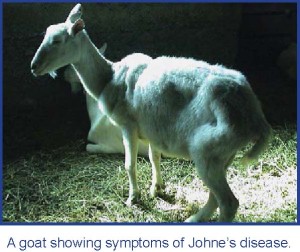
A: A goat that appears perfectly healthy can be infected with MAP.Although goats become infected in the first few months of life, many remain free of clinical illness until months or years later. When goats finally do become ill, the symptoms are vague and similar to other ailments: rapid weight loss and, in some cases, diarrhea. Despite continuing to eat well, infected goats soon become emaciated and weak.
Since the signs of Johne’s disease are similar to those for several other diseases—parasitism, dental disease, Caseous Lymphadenitis (CLA) and Caprine Arthritis-Encephalitis Virus (CAEV)—laboratory tests are needed to confirm a diagnosis.
When an animal with signs of Johne’s disease is discovered, it is very likely that other infected animals—even those that still appear healthy— are in the herd. Control of the infection requires that you and your veterinarian address it in the whole herd and not just on an individual animal basis.
Q: Why do goats with clinical signs of Johne’s disease lose weight and become weak?
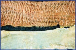
A: When an animal is infected with MAP, the bacteria reside in the last part of the small intestine—the ileum—and the intestinal lymph nodes. At some point, the infection progresses as bacteria multiply and take over more and more of the tissue. The goat’s immune system responds to the bacteria with inflammation that thickens the intestinal wall and prevents it from absorbing nutrients. As a result, a goat in the clinically ill stage of Johne’s disease in effect starves to death. At this stage the organism may also spread beyond the gastrointestinal tract, travelling in the blood to muscles or other major organs such as the liver or lungs.
Top: Thickened intestinal mucosa caused by Johne’s disease.
Bottom: Thin, pliable, normal intestine
Q: How do goats become infected? How is MAP spread in a herd?
A: Johne’s disease usually enters a herd when an infected, but healthy-looking, goat is purchased. With MAP hiding in its small intestine, this infected goat sheds the organism in its pellets and into the environment— perhaps onto pasture or into water shared by its new herdmates. Goats are at risk when they repeatedly swallow the organism, especially when they are young (less than 6 months old). If the doe is infected, her offspring can become infected even before they are born (in utero transmission). The organism is also shed in an infected doe’s milk and colostrum.
Kids are most susceptible to infection with MAP and often become infected through ingestion of manure containingMAP— such as from suckling manure-stained teats, swallowing milk that carries MAP or eating feed, grass or water containing MAP-contaminated manure. Bottle-fed kids can also become infected if the milk was contaminated. Heat treatment used to control CAE in milk is not sufficient to kill MAP organisms.
Since goats usually produce more than one kid per birthing, Johne’s disease can spread swiftly through a herd, especially if the infection remains undetected for several kidding seasons. While there seems to be age-related resistance to Johne’s disease, some older goats may become infected, particularly when their immune systems are suppressed for other reasons. Johne’s disease can be transmitted from one ruminant species to another—for example from cows to goats, goats to sheep, etc.
Q: When do infected animals shed the bacteria?
A: Infected goats shed MAP in their manure on and off throughout their lives. The older the animal, the more likely that shedding occurs as the infection progresses. As goats enter the latter stages of infection and clinical signs begin to appear, the infective organism is shed more often and more heavily.
Q: Is it difficult to find out if my herd has Johne’s disease?
A: Sometimes. Johne’s disease is often mistaken for intestinal parasitism, chronic malnutrition, environmental toxins, cancer and CLA— particularly in goats that have internal abscesses. Many herds rotate parasite treatments for several rounds before testing for Johne’s disease and determining this is the reason their goats are so thin. In addition, some of the common laboratory tests for Johne’s disease may be difficult to interpret.
If Johne’s disease is suspected but has not been confirmed in a herd, a necropsy of a goat with symptoms of the disease may be helpful in determining if the infection is in a herd. This necropsy may reveal enlarged intestinal lymph nodes and a thickened, corrugated intestinal tract.
A complete necropsy of a goat suspected of having Johne’s disease should include culture of the intestine and adjacent lymph node as well as microscopic examination of these tissues to give you the greatest confidence in the diagnosis.
Q: How can I help keep Johne’s disease out of my herd?
A: Buyers beware! The most common way that the infection is introduced to a herd is through the purchase of an animal from an infected herd. Since many people raising goats are unaware of Johne’s disease, both the seller and buyer are usually shocked when the diagnosis is made.
In short, it is easier to keep MAP out of a herd than to control the disease once MAP sneaks in.
Practices that can help prevent the introduction of Johne’s disease into a herd are:
- Maintain a closed herd. Don’t buy Johne’s disease.
- If you bring animals into the herd, purchase animals only from herds that have tested for Johne’s disease. Ideally, purchase only from herds that have a negative whole-herd test in the last year. If this is not possible, you are better off buying from someone who is aware of the infection, has tested for it and can provide accurate records on the disease in their herd than to purchase an animal from someone who has never heard of Johne’s disease.
- If no diagnostic testing has been conducted in the source herd, at least closely evaluate the body condition of all the adult animals, discuss the history of clinical signs in any animals in the herd over the past few years with the seller and test the adult animal to be purchased.
- If the animal to be purchased is less than a year old, test its dam since infected young animals are unlikely to test positive for the infection.
- Avoid grazing goats on pastures where MAP-infected ruminants have grazed. Graze young goats on such a pasture only after it has rested for a year. To date, MAP infection of free-ranging ruminants such as deer or elk is uncommon, and currently these species are not believed to be an important source of infection to farmed ruminants.
- Do not bring in other species that are susceptible to Johne’s disease: sheep, cattle, other ruminants.
- Do not board or borrow other people’s goats, as this can introduce the infection into your herd.
Q: How can I control Johne’s disease once it has entered my goat herd?
A: Since there is no cure for Johne’s disease, control of the infection is critical. Control of Johne’s disease takes time and a strong commitment to management practices focused on keeping young animals away from contaminated manure, milk, feed and water. A typical herd clean-up program may take a number of years. The basics of control are simple: new infections must be prevented, and animals with the infection must be identified and removed from the herd.
Your State Designated Johne’s Coordinator can help you undertake an on-farm risk assessment that evaluates your operation, your resources and your goals. An on-farm risk assessment highlights current management practices that may put your herd at risk for spreading Johne’s disease and other infections. At the completion of a risk assessment, your veterinarian can work with you to develop a management plan designed specifically for you and your herd that will minimize the identified risks for disease transmission. (An on-farm risk assessment is part of the Johne’s disease course for goat producers at www.vetmedce.org.)
Most control plans follow basic rules of sanitation to block transmission of the infection within the herd. Management recommendations include:
- Keep kidding areas as manure free as possible. Use deep, fresh bedding or sunny pastures with minimal manure.
- If feasible, clean the udders of dams before kids nurse.
- Use milk and colostrum from test-negative animals.
- Be aware that colostrum purchased from another goat herd or cow herd may be contaminated. Pasteurization needs to be at 145°F (63°C) for 30 minutes for batch pasteurization, or 162°F (72°C) for 15 seconds for flash pasteurization to kill MAP in milk.
- Kid suspect or test-positive does in an area separate from the test-negative does.
- Move young animals and their mothers to “clean” pastures as soon as possible after kidding.
- Wean early and put young goats on uncontaminated pastures.
- Keep water sources clean, particularly those used by kids. Use waterers designed to minimize fecal contamination.
- Raise all feeders and avoid feeding on the ground.
- Use diagnostic tests to identify infected animals and remove them from the herd.
- Necropsy sick or cull animals to determine if your herd is infected with MAP.
- If your herd has had numerous cases of Johne’s disease, discuss depopulation with your veterinarian or, at a minimum, immediately remove all test-positive animals and their last-born kid. Do not allow kids to be exposed to milk or manure from infected animals.
Remember: Preventing Johne’s disease is much less costly than controlling it.
Q: How can I clean contaminated equipment, stalls and fields?
A: The MAP organism is very hardy in the environment: It resists heat, cold, drying and dampness. Although the majority of organisms die after several months, some will remain for many months. In fact research shows that MAPcan survive—at low levels—for up to 11 months in soil and 17 months in water. MAP has also been recovered from grasses fertilized with MAP-contaminated manure. This is why pastures and fields known to be contaminated with MAPshould not be used for kids, calves or lambs for at least one year after last exposure.
Feed and watering equipment that may have become contaminated should be washed and rinsed. When cleaning a water trough, sediment and slime from the sides and bottom should not be dumped onto ground that will be grazed by young goats as the sediment and slime may be contaminated with MAP.
Disinfectants labeled as “tuberculocidal” may be used as directed for cleaning tools, implements and some surfaces. These disinfectants, however, can be inactivated by organic material—such as dirt and manure—and therefore are not effective on dirty surfaces, wood surfaces, soil or even cement floors.
Composting of manure and used bedding can reduce the number of living MAP organisms.
Q: Should I test my herd for Johne’s disease?
A: If you have goats with a normal appetite that have become thin and are not responding to treatment, talk to your veterinarian. The culprit may be Johne’s disease.
Remember: Since Johne’s disease is a herd problem, testing should focus on the herd and not just a single animal.
Diagnostic testing for Johne’s disease can help to:
- Determine if MAP infection is present in your herd.
- Estimate the extent of MAP infection in your herd.
- Control MAP in an infected herd.
- Make a diagnosis for a sick animal.
- Check if MAP is present in the environment.
- Meet a pre-purchase or shipping requirement.
- Demonstrate to potential buyers that your animals are low risk for Johne’s disease (test-negative). Once your veterinarian knows your goals in testing for Johne’s disease, a testing plan that best meets your needs can be put in place. This plan should outline the type of test, when to test, which animals to focus on, the cost of testing, how to interpret the results and what actions to take based on test results. Decide how you plan to use your test results before you collect the samples.
Q: What diagnostic tests are available? Which one is best?
A: Although there is no one “best test” for Johne’s disease in goats, the best testing plan is one developed by you and your veterinarian since you know your operation best—its goals, resources, other animal health issues.
Diagnostic tests for Johne’s disease look for either the organism that causes Johne’s disease (MAP) or the animal’s response to infection.
Tests that look for the organism include culture and PCR, with manure samples tested. Individual animals can be tested or a laboratory can pool manure samples from multiple animals and provide owners with effective Johne’s disease surveillance for a fraction of the cost of individual culture or PCR.
The animal’s body responds to infection by making antibodies. Tests that measure antibody levels are the ELISA for milk and blood samples. Due to the biology of MAP infection, older goats are much more likely to shed MAP or produce antibody. Therefore, diagnostic tests are less reliable for goats that are less than 18 months old.
Testing approaches that have worked well for other goat herds include:
| Testing Purpose | Option A | Option B |
|---|---|---|
| Confirm presence of MAP in a herd. | Culture 5 – 10 environmental fecal samples collected at high goat traffic areas on the farm. | Using ELISA* or fecal culture, test the oldest or thinnest goats— 10% or more of the herd. |
| Determine number of goats that are infected. | Blood test (ELISA*) all adult goats. | Collect fecal samples for the lab to test by pooling for culture. For every positive pool, samples are retested individually. |
| Control or eradicate MAP in an infected herd. | Blood test (ELISA*) goats after their second kidding or older. | Collect fecal samples for the lab to test by pooling for culture. For every positive pool, samples are retested individually. |
| Diagnose a sick goat or goat with weight loss and diarrhea. | If previous cases have been seen in the herd: ELISA.* (Fecal culture if CLA is a problem in the herd or if the herd has been vaccinated for CLA.) | If MAP has never been confirmed in the herd, use fecal culture. *Use commercial ELISA kit approved by the USDA for small ruminants to limit the chance of false-positive results due to cross-reacting antibodies from other types of infections. Test samples should be submitted to a laboratory that has passed an annual “check test” demonstrating their competency. These labs are listed here. |
Q: Where can I find more information about Johne’s disease?
A: The University of Wisconsin School of Veterinary Medicine’s Johne’s disease website—www.johnes.org—addresses all aspects of Johne’s disease for multiple species, including goats. The site has an “Ask An Expert” feature that allows you to submit your own questions and receive a personalized response from an expert.
The University of Wisconsin School of Veterinary Medicine also offers a free online course for goat producers. Simply go to www.vetmedce.org, click on “Courses” in the lower left hand corner of the homepage. Once on a new page, click on “Johne’s Disease.” At the next new page, click on “Johne’s Disease Courses for Producers” followed by clicking on “0017—Johne’s Disease for Goat Producers.”
To learn more about Johne’s disease in goats, please contact your State animal health regulatory agency or your State Designated Johne’s Coordinator. Contact information for your State’s Johne’s disease program is available online at www.johnesdisease.org when you click on “State Contacts.”
